chroogomphus
Posted by Fred 11/21/12 - - - - - - Scroll Down to See the Discussion
This is a very confusing series of posts.
There were several different posts and apparently some emails
that are separate from the posts.
Clearly some of the messages exchanged could not be included in the following since they
were not on the "What is this mushroom site"
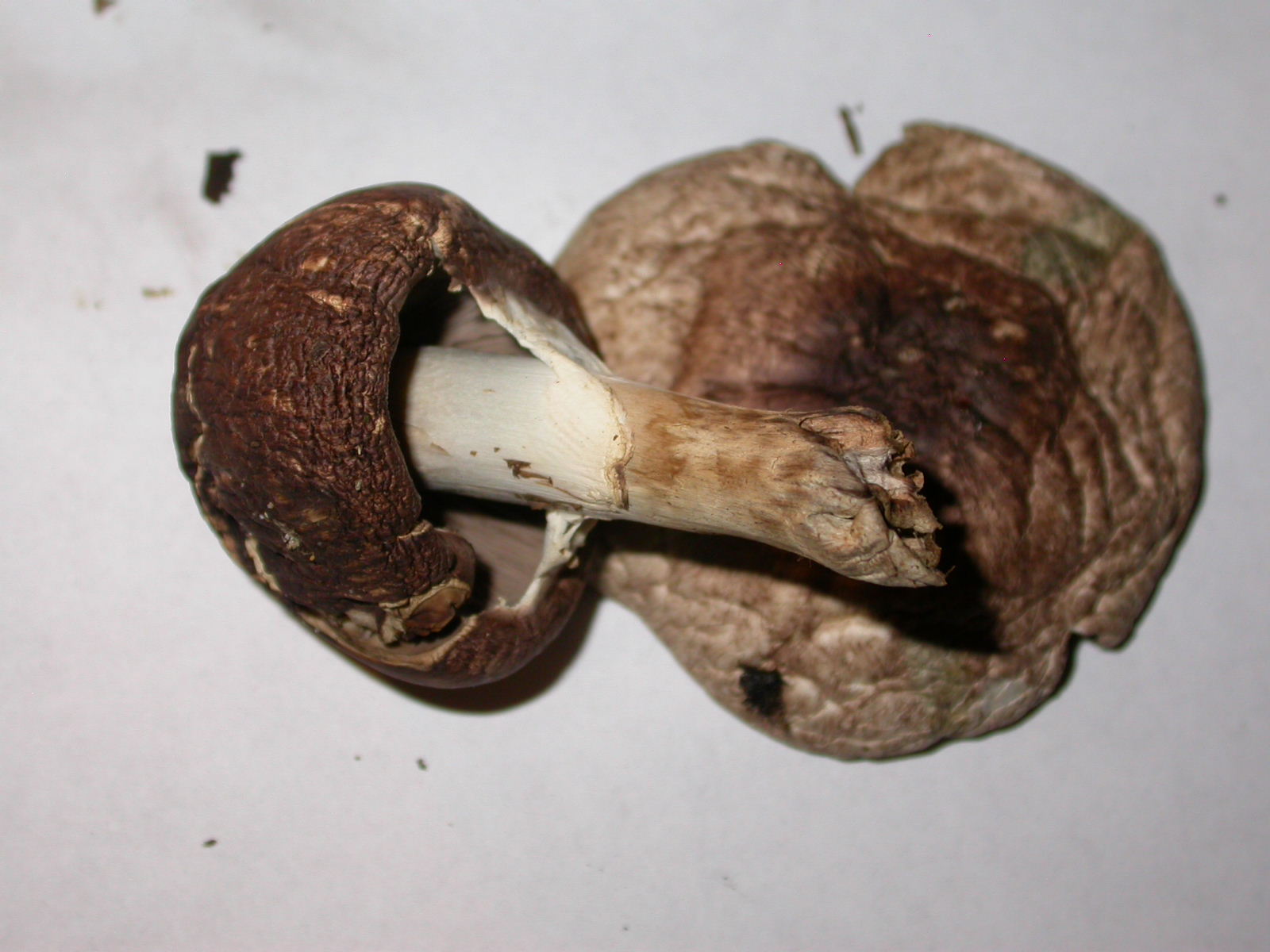
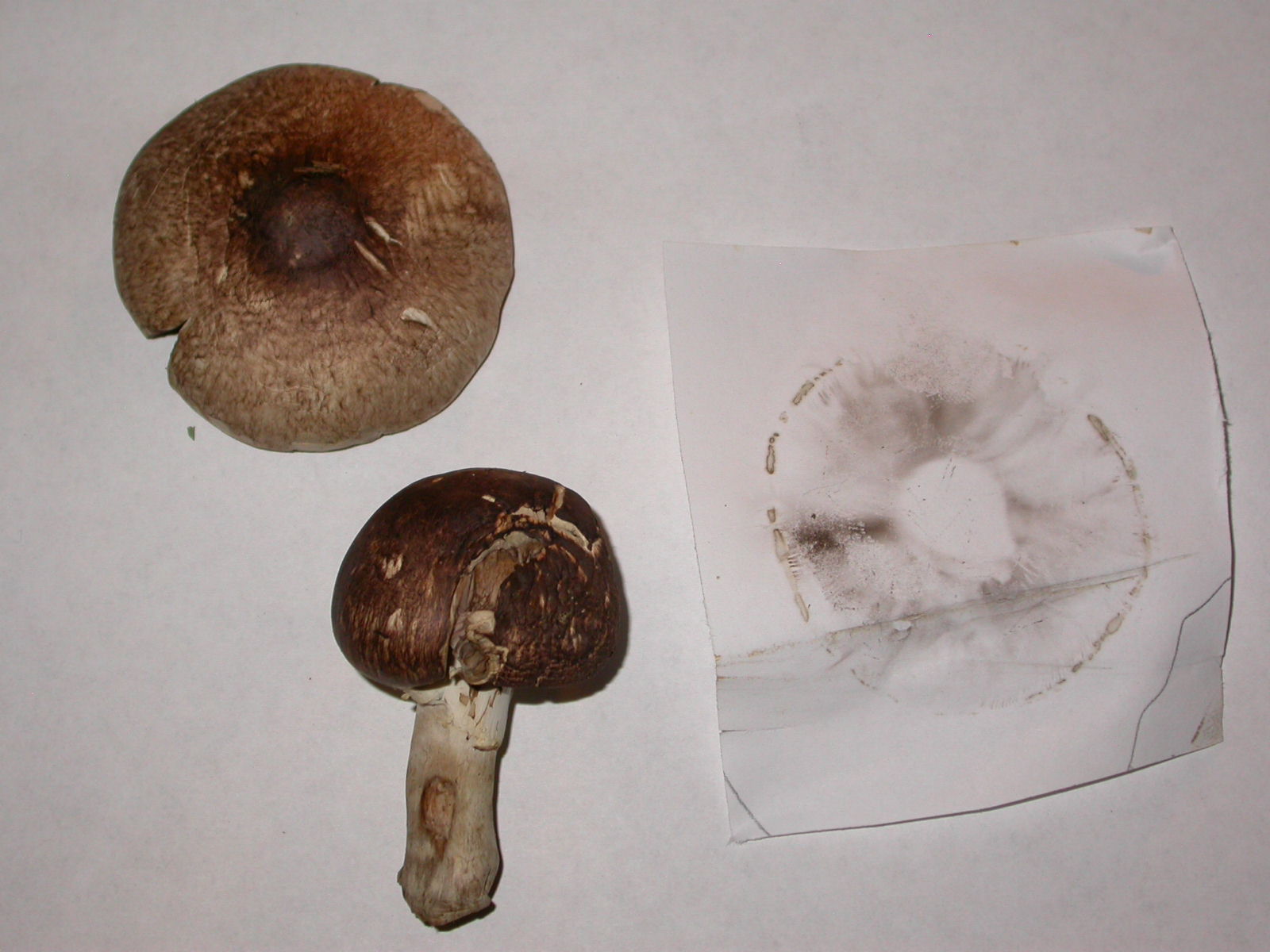
Fred: Under deep conifer cover and duff at park at coast
lost base of large specimen by mishandling and had to throw out older specimen cause too many "critters"....
initially thinking Agaricus subrufescens but discouraged by Matchmaker description of range and occurence
Stipe not broad as I would expect.
KOH on cap looked like it left a faint yellow olive green
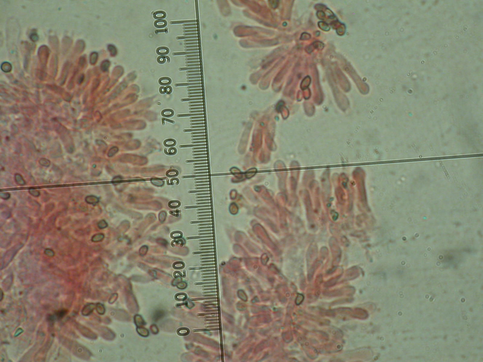
..44 image
400x 1.25 micron per unit
note basidia left of 60-80 marks both 2 pronged and 4 pronged
h20 washed after phloxine and congo red flushes
---------- It appears that a post by Fred was lost ----
Fred: cystidia approximately the same? yes.
cuticle hyphae width should be (???) From Matchmaker:
C. vinicolor
cap cuticle of hyphae 6-7 microns wide,
appressed, gelatinous, colorless, light yellow-brown in KOH, scattered weakly amyloid hyphae;
C. ostraceous
cap cuticle a pellicle of gelatinized hyphae,
(2.4)5-7.5(8.4) microns wide, "hyaline in KOH, hyaline in Melzer's solution,
in crushed mounts hyaline to yellow with many yellow granules within"
attached photo 6358 of pellicle hyphae:
.jpeg) 400x 1.25 micron
400x 1.25 micron
Lugol's solution
Photo 6360 -trama hyphae show weakly amyloid reactions
200x 2.5 micron/div
lugols solution
Fred: gonna cost me $50.00 to get the paclink address into my mailer....
so I'm sending onto you to post WTM if you so desire...thx
thickwalled or thinwalled cystidia?
DSCN6335 @ 400x 1.25 microns to unit Congo red washed w h2O
C tomentosus
spores 15-25 x 6-9 microns, narrowly elliptic to spindle-shaped, smooth,
cystidia on gills 118-255 x 10-21 microns, cylindric to fusiform,
with fairly thick walls 2-4 microns at thickest part,
occasionally thin-walled, sometimes containing yellow-brown granules in KOH
C vinicolor (our vote 'cept for cystidia wall thickness confusion)
spores (14)17-23 x 4.5-7.5 microns, more or less spindle-shaped, smooth
gill cystidia 112-164 x 13-19.5 microns,
fusoid-ventricose, narrowly clavate to narrowly fusiform,
thick-walled (walls reaching 5-7.5 microns at thickest part),
thickening toward middle of cystidium,
the thickened wall light to dark amyloid in some cystidia but in KOH colorless to yellow brown
significant cystidia length differences.
Sava: - Forwarded message From: Fred ..@gmail.com> Date: Tue, Nov 20, To: Sava Krstic >
gonna cost me $50.00 to get the paclink address into my mailer.... so I'm s
I would eliminate C. tomentosus based on the gill and stem color,
which are both orange in this species (though gill become dark with age).
To me, the real dilemma is: C. vinicolor or C. rutilus/C.ochraceus.
I am not confident with judging thick- or thin-walled (would bet thin-walled here),
but am hoping that Judy will help us here.
Looking at the spore sizes,
it seems that the spores in C. vinicolor are more elongated than those in C. rutilus
and thus closer to what your photo shows.
Thus, if I had to guess, I'd say your mushroom is C. vinicolor.

 Dick B: Based on one cystidia I would agree with your conclusion.
Are the majority of the cystidia this shape and thickness?
If so I would suggest checking the cuticle hyphae width to confirm your ID.
Dickj B: I agree that the description of the microscopic specifics is very confusing.
The reason is that Miller at some point separated C. rutilus and C. ochraceus
so Ian for some reason decided to include the description for both C. ochraceus and C. rutilus
under C. ochraceus in MatchMaker. Bottom line the first description is for C. ochraceus.
That's the one you are interested in. C. rutilus is considered a European species.
The cuticle hypae for C. ochraceus is 3-6u.-
dick B: I'm not sure who to reply to but thought one of you could post if appropriate.
I spent some time trying to understand Chroogomphus at a fall foray a few years ago.
The part of the cystidia you are seeing is thin walled.
If you squash your section and break out a full cystidia you will probably
find that the middle and lower part of the cystidia is thick walled. Also will come close to
doubling the length of the cystidia and closer to specified length. Hope this helps.