gymnopilus or tricholompsis
Posted by L&J N 11/11/12 - - - - - Scroll Down to See the Discussion
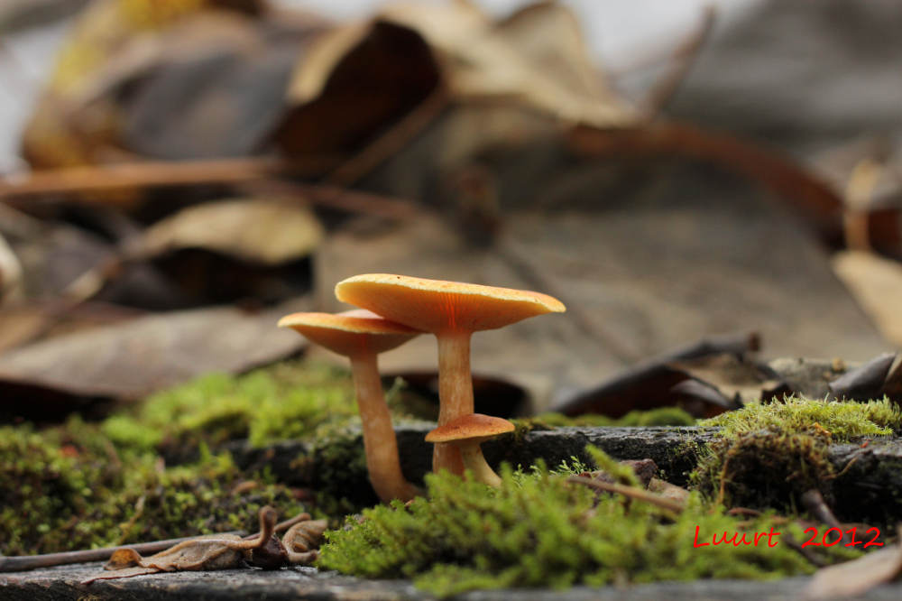
LJ& N: This was growing on some old rotten wood (plywood, 2 by 4).
The cap of the larger specimen is about 3 inches across.
spores white
The first guess was Tricholompsis decora
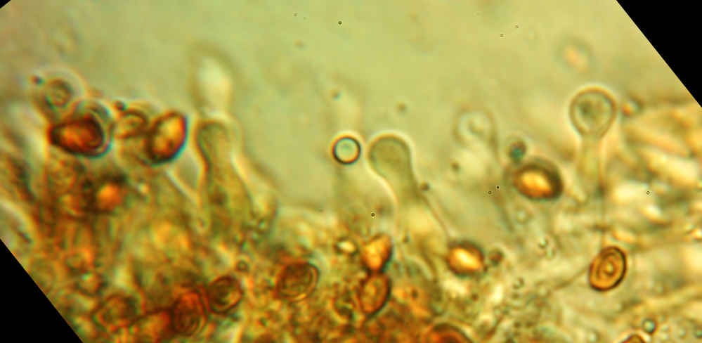
but as you can see from the last (poor) image, the spores are NOT smooth.
Where do I look next to identify this?
Sava: Interesting! I'm not sure the last picture proves that the spores are not smooth.
It looks like there are veil remnants on the upper part of the stem.
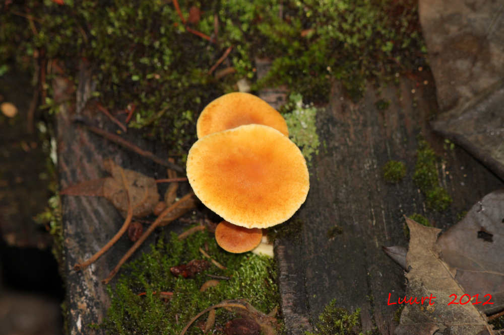
Are the gills yellow?
Judy: In looking closely at enlargements of your photos –
did you really get a definitely white sporeprint?
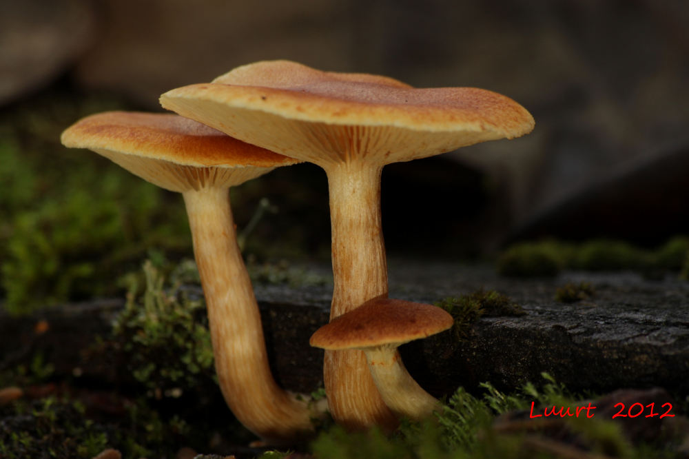
The smallest specimen looks to have veil remnants that are brownish,
and the back specimen looks to have a faint trace of veil fibers on the stipe as well.
Also, on that back specimen, look closely at the cap surface under the gills
of the overtopping one – one could argue for some brown spores too. With it growing on woody material, and that inately fibrillose cap and the
fibrillose cap and stipe and the spores that look like they could be
slightly roughened (would need slightly clearer views of them),
it resembles A Gymnopilus more than a Tricholomopsis.
Tricholomopsis has smooth white spores, and if you lay one gill on it’s side in water,
place the coverslip on it and tap gently to make it thinner and spread it
out a little (not a lot!) – are there cystidia on the gill edges?
Describe or put on a pic of them:
are they somewhat fat and inflated with thin walls or are they thinnish,
with a knobby head and thin neck?
that should tell us all a lot more.
LJ&N : "Tricholompsis or gymnopilus?
Judy R asked: did you really get a definitely white sporeprint?"
No, and that's an "oops" on my part.
I tried to get a spore print while the sample was still on location outside,
and I only got a thin print.
It looked both white spored as well as roughened spores.
I just collected a 3 day sampling on a microscope slide (still on-location outside)
and the spore print is pale yellow-ochre
If you create a color swatch in Photoshop,
the colors are approximately Red=245 Green=220 Blue=100.
Possibly a very light rusty-orange.
The stipe is definitely fibrilose,
Spore size, if I've done my calibrations right, are about 3 by 5 microns.
"Judy asked: are there cystidia on the gill edges?
Describe or put on a pic of them:
are they somewhat fat and inflated with thin walls or are they thinnish,
with a knobby head and thin neck? that should tell us all a lot more."
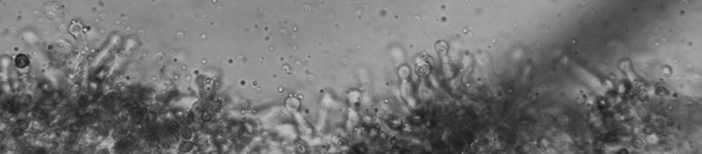
It seems like I can see both, depending on where I look
Some are sausage-shaped, a few are more like bowling pins.
Sorry about the image quality.
An Inverted Stage microscope is not very amenable to oil immersion techniques.
As it is I have to seal the cover slip on because the close
focusing distances require the slide to be placed on the stage cover slip side down.
I'm at a loss to explain why my early attempts at viewing the spores produced rough surfaces,
while this latest spore samples produced relatively smooth spores.
Could it that be because the first ones were done in air while the later smooth ones were
done under a water / coverslip?